

As name indicates these technique imparts different colour to different bacteria or bacterial structure. It allows us to differentiate between different kinds of bacterial cells or different parts of a bacterial cell. Differential stain technique includes the following technique:-
2. Acid Fast staining/Zeihl-Neelsen staining
1. Gram staining
The most commonly used differential stain is the Gram stain, first described in 1884 by Christian Gram.
The Gram stains divides bacteria into two groups, i.e. Gram-positive and Gram-negative. Those organisms which retain the primary stain (crystal violet) are stained purple and are designated Gram-positive; those which lose the crystal violet and are subsequently stained by a safranin counterstain appear red and are designated Gram-negative.
Procedure of gram staining
- First, crystal violet, a primary stain, is applied to a heat-fixed smear, giving all of the cells a purple color.
- Next, Gram’s iodine, a mordant, is added. A mordant is a substance used to set or stabilize stains or dyes; in this case, Gram’s iodine acts like a trapping agent that complexes with the crystal violet, making the crystal violet–iodine complex clump and stay contained in thick layers of peptidoglycan in the cell walls.
- Next, a decolorizing agent is added, usually ethanol or an acetone/ethanol solution. Cells that have thick peptidoglycan layers in their cell walls are much less affected by the decolorizing agent; they generally retain the crystal violet dye and remain purple. However, the decolorizing agent more easily washes the dye out of cells with thinner peptidoglycan layers, making them again colorless.
- Finally, a secondary counterstain, usually safranin, is added. This stains the decolorized cells pink/red and is less noticeable in the cells that still contain the crystal violet dye.

| Steps | Procedure | Effect/outcome |
|---|---|---|
| Primary stain(crystal violet) | Add several drops of crystal violet to the smear and allow it to sit for 1 minute. Rinse the slide with water. | Both Gram-positive and Gram-negative cells will be stained purple by the crystal violet dye |
| Mordant (iodine) | Add several drops of iodine to the smear and allow it to sit for 1 minute. Rinse the slide with water. | Iodine as mordant increases the affinity of the cells for the crystal violet |
| Decolorization (ethanol/acetone) | Add drops of ethanol one at a time until the runoff is clear. Rinse the slide with water. | Gram-positive cells resist decolorization and remain purple. The dye is released from Gram-negative cells |
| Counterstain(safranin) | Add several drops of safranin to the smear and allow it to sit for one minute. Rinse the slide with water and blot dry. | Gram-negative cells stain by the safranin and become pink |
Principle of Gram Staining
During the decolorization step, alcohol may extract the lipids, increasing the porosity or permeability of the cell walls. Thus, the crystal violet-iodine complex is easily lost. The Gram-positive bacteria, however, do not have lipid-rich cell walls. Their cell walls become dehydrated during the alcohol treatment, decreasing the porosity so that the crystal violet-iodine complex is retained.
2. Acid Fast staining/Zeihl-Neelsen staining
It is the differential staining technique that was first developed by Ziehl and later on modified by Neelsen. So this method is also called Ziehl-Neelsen staining techniques. Neelsen in 1883 used Ziehl’s carbol-fuchsin and heat then decolorized with acid alcohol and counter-stained with methylene blue.
It is used to differentiate bacteria into an acid-fast groups and non-acid fast groups. This method is used for those microorganisms which are not staining by simple or Gram staining method, particularly the member of genus Mycobacterium, are resistant and can only be visualized by acid-fast staining.

Principle of Acid-Fast Stain
When the smear is stained with carbol fuchsin, it solubilizes the lipoidal material present in the Mycobacterial cell wall but by the application of heat, carbol fuchsin further penetrates through lipoidal wall and enters into cytoplasm. Then after all cell appears red. Then the smear is decolorized with decolorizing agent (3% HCL in 95% alcohol) but the acid fast cells are resistant due to the presence of large amount of lipoidal material in their cell wall which prevents the penetration of decolorizing solution. The non-acid fast organism lack the lipoidal material in their cell wall due to which they are easily decolorized, leaving the cells colorless. Then the smear is stained with counterstain, methylene blue. Only decolorized cells absorb the counter stain and take its color and appears blue while acid-fast cells retain the red color.
Procedure of Acid-Fast Stain
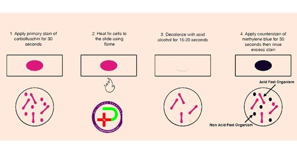
- On a clean sterile microscopic slide, make the smear of the sample culture and heat fix the smear over blue heat.
- Over the smear, pour and flood the smear with carbol fuschin and heat gently until it produces fumes.
- Allow it to stand for 5 minutes and wash it off with gently flowing tap water.
- Add 20% sulphuric acid and leave it for 1-2 minutes. Repeat this step until the smear appears pink in color.
- Wash off the acid with water.
- Flood the smear with methylene blue dye and leave it for 2-3 minutes and wash with water.
- Air dry and examine the stain under the oil immersion lens.
| Application of | Reagent | Cell colour | |
| Acid-fast | Non-acid fast | ||
| Primary dye | Carbol fuchsin | Red | Red |
| Decolorizer | Acid alcohol | Red | Colorless |
| Counterstain | Methylene blue | Red | Blue |
3. Albert’s stains
Its application aim at identifying bacteria that contain special structures known as metachromatic granules. Other staining techniques that are used to detect the presence of granules in the cytoplasmic membrane of bacteria are Nessers’s stain and Pugh’s stain.
Albert stain is made up of two staining solutions; designated as Albert Solution 1 and Albert Solution 2.
Albert Solution 1:toluidine blue, malachite green, glacial acetic acid, and alcohol
Albert solution 2:Iodine and Potassium iodide in water
Albert staining solution 1 acts as the staining solution while Albert solution 2 acts as the mordant, i.e an ion element that binds and holds a chemical dye, to make it stuck on the micro-organism.
Procedure
A. Staining:
- Aseptically, take a loopful culture of Corneybacterium diphtheriae
- Make a smear at the center of a clean sterile glass slide
- Heat fix the smear, gently
- On a staining rack, place the smeared glass slide.
- Add Albert staining Solution 1 into the smear and leave it for 3-5 minutes
- Wash the smeared slide with gently flowing tap water
B. Mordanting
- Add Albert staining solution 2 and leave it for 1 minute
- Wash the slide with gently flowing tap water.
- Blot to dry the smeared glass slide
- Add cedarwood oil on the smear
- Then observe under a microscope by oil immersion at 1000x
The metachromatic granules stain bluish black while the rest of the microbial cell stains green.
Differential staining MCQ with Answer
Differential staining FAQ
1. Compare simple staining and differential staining?
- Simple Staining-
- imparts same colour to all the bacteria in smear
- uses only one dye
- helps to observe the size, shape, structure and arrangement of bacterial cells.
- Example with Thailand blue and basic fuchsin dye.
- Differential Staining-
- imparts different colour to different bacteria or bacterial structure
- uses more than one dye.
- It imparts two or more different colours to the microbial cells
- helps to differentiate between different types of bacterial species.
- Example- Gram’s staining, acid fast staining & Albert staining.
2. What is difference between simple and differential staining?
Simple stain imparts same colour to all the bacteria in smear. Example- Methylene blue staining
Differential staining imparts different colour to different bacteria or bacterial structure. Example- Gram’s staining, acid fast staining & Albert staining.
3. What is the advantage of differential staining ?
- It gives quick results when examining infections (to find gram positive or negative bacteria)
- It is simple and cost-effective.
- It helps with determining appropriate treatments for infection. whether an infection is caused by bacteria, fungi or viruses,
- It is basically a key procedure in identifying bacteria
4. What is the purpose of mordant in differential staining?
Mordant increases the affinity of the cells for the primary dye/crystal violet. Gram’s iodine acts as a mordant (Helps to fix the primary dye to the cell wall). Iodine makes the protoplasm more acidic. which account for retaining the basic primary stain. Iodine also combine with dye to form a dye-iodine complex and fixes the dye on bacterial cell. Gram positive cell wall or cytoplasmic membrane is less permeable, dye-iodine complex gets trapped within the cell of gram + bacteria. While Gram negative cell wall has increased permeability when subjected to acetone/alcohol, result into outflow of dye-iodine complex during decolorization step.
5. What is the purpose of decolorizing agent in differential staining?
Decolorizer is used to remove the primary stain (crystal violet) from Gram Negative bacteria (those with LPS imbedded in their cell walls). Decolorizer is composed of an organic solvent, such as, acetone or ethanol or a combination of both.)
Gram-positive organisms are able to retain the crystal violet stain because of the high amount of peptidoglycan in the cell wall. Gram-positive cell walls typically lack the outer membrane found in Gram-negative bacteria.
9. What is the purpose of counterstain in differential staining procedure?
A counter stain (Safranin), is applied to stain those cells (Gram Negative) that have lost the primary stain as a result of decolorization
Comments are closed